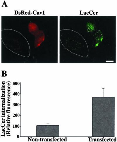
Figure 7.
Caveolin-1 overexpression stimulates LacCer internalization. HeLa cells were transfected with DsRed-cav-1 for 48 h. Cells were then pulse-labeled with BODIPY-LacCer as in Figure 4. (A) Fluorescence micrograph showing two transfected cells (identified by DsRed fluorescence) and a nontransfected cell in the same field. LacCer was observed at green wavelengths (see MATERIALS AND METHODS). Note the stimulation of LacCer internalization in the transfected cell, relative to the neighboring nontransfected cell outlined in white. Bar, 10 μm. (B) Quantitation of LacCer internalization in transfected vs. nontransfected cells by image analysis. Values are the mean ± SD of at least 10 cells in each of three independent experiments.

