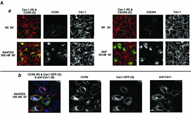
Figure 9A.
Caveolin-1 and -3 differentially regulate agonist-dependent endocytosis of CCR5 and CXCR4. HeLa cells were transfected with the indicated chemokine receptor and treated with the indicated agonists. Cell surface–bound antibody was stripped by acid wash. Color overlays and individual channels are laid out left to right. R, red; G, green; B, blue. (a) Endocytic trafficking patterns of agonist-occupied CCR5 and CXCR4 in HeLa cells stained for endogenous caveolin-1. Receptor endocytosis was visualized by antibody feeding using APC-conjugated 1o mAbs (pseudocolored green). Caveolin-1 was detected by staining with rabbit anticaveolin-1 IgG followed by 2o staining with Alexa 488–conjugated anti-rabbit IgG (red). (b) CCR5 trafficking in HeLa cells overexpressing caveolin1-GFP. Agonist treatment was restricted to 45 min at 100 nM to limit CCR5 endocytosis. CCR5 trafficking was visualized by feeding unconjugated 3A9 mAb. Internalized antibody was visualized by staining with Alexa 568–conjugated anti-mouse IgG. Fluorescence from exogenous Cav1-GFP is in green. Both endogenous and plasmid-expressed caveoin-1 were detected by staining with rabbit IgG against caveolin-1 followed by staining with Alexa 647–labeled anti-rabbit IgG as the 2o reagent. Note that Cav1-GFP fluorescence and Cav1 antibody staining do not overlap completely, presumably because the epitope recognized by rabbit antibody is not exposed on all caveolin molecules.

