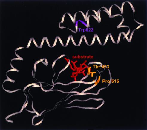
Figure 5.
Mutations in kar2-1 and kar2-133 reside in the peptide-binding domain of BiP. The yeast BiP and E. coli DnaK amino acid sequences were aligned and mutated residues in kar2-1 (P515L) and kar2-133 (T473F) were mapped onto the three-dimensional structure of the DnaK peptide-binding domain (Zhu et al., 1996). The mutated residues are highlighted in yellow, and the approximate location of the lone tryptophan (at position 622) in yeast BiP is indicated in magenta. The bound substrate is displayed in red and is oriented perpendicular to the page.

