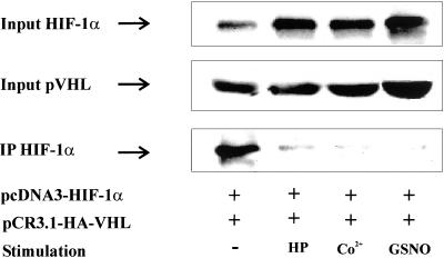
Figure 6.
In vivo interaction between HIF-1α and pVHL. HEK293 cells were cotransfected with 1 μg pCR3.1-HA-VHL and 3 μg pcDNA3-HIF-1α expression plasmids. After changing medium at 16 h, incubations continued for 8 h before stimulation with 1% hypoxia (HP), 100 μM CoCl2, or 1 mM GSNO for 4 h, followed by MG132 for 1 h as described in MATERIALS AND METHODS. Coimmunoprecipitation was performed with an anti-HA mAb, which recognized heme agglutinin-tagged pVHL and anti-mouse antibody-coated magnetic beads. Expression of pVHL (input) and accumulation of HIF-1α in cell lysates (input) as well as immunoprecipitates (IP) was determined by Western analysis using an anti–HIF-1α mAb as described in MATERIALS AND METHODS. Each experiment was performed at least three times and representative data are shown.

