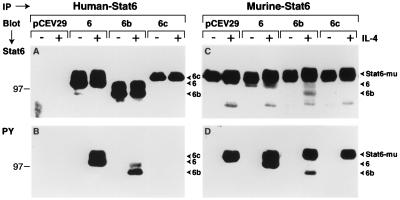
Figure 3.
Expression and IL-4-induced tyrosine phosphorylation of Stat6 isoforms in FDC-P2 cells. (A and B) Detection of human Stat6. FDC-P2 transfectants were starved as indicated and then stimulated with IL-4 (500 ng/ml) for 20 min. Whole cell lysates containing equivalent amounts of protein were then immunoprecipitated (IP) with anti-human Stat6 serum and subjected to SDS/PAGE. Resolved proteins were transferred to Immobilon-P membranes and immunoblotted (Blot) with anti-human Stat6 serum (A) or anti-phosphotyrosine (PY) (B) (26). (C and D) Detection of mouse/human Stat6. FDC-P2 or FDC-P2–Stat6 isoform transfectants were treated with IL-4, immunoprecipitated with anti-Stat6 serum, and subjected to SDS/PAGE. Immobilon-P membranes were immunoblotted with anti-Stat6 serum (C) or anti-phosphotyrosine (D). Bound primary antibody was detected by anti-rabbit or anti-mouse antibody conjugated to horseradish peroxidase followed by enhanced chemiluminescence (Amersham).

