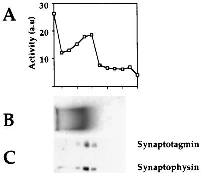
Figure 3.
Agarose gel electrophoresis of peak II vesicle membranes. Peak I and II membranes were pelleted and electrophoresed in a 0.15% agarose gel in 50 mM Mes buffer (pH 6.5) for 24 h at 0.75 V/cm while recirculating the Mes electrophoresis buffer at 4°C (23). The lane containing peak I was stained with Coomassie Blue for direct visualization (B), and the peak II-containing lane was cut into fractions and incubated with synthetic substrate to localize the aminopeptidase activity (A). An aliquot from each fraction was electrophoresed in a 10% SDS/PAGE gel, and Western blot analysis was performed with anti-synaptotagmin and anti-synaptophysin antibodies (C).

