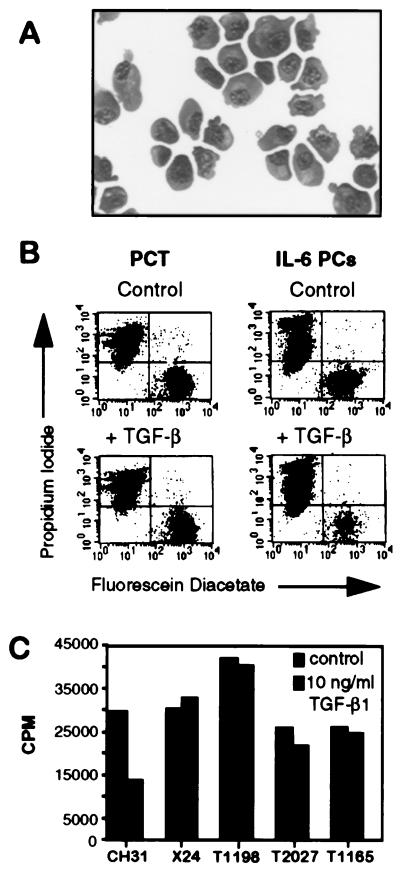
Figure 1.
(A) Stained cytofuge preparation of purified plasma cells from an IL-6 transgenic mouse. Pooled lymph nodes were perfused, and cells were incubated at 37° to remove adherent cells. The remaining cell suspension was depleted of B220+ and Thy1.2+ cells by using MACS. (B) FACS analysis of cell viability in response to treatment with TGF-β. Comparison of MACS-purified, untreated primary PCT cells (Upper Left) with those treated with 1 ng/ml TGF-β (Lower Left) shows that TGF-β has no effect on the viability of PCTs after 48 h. The percentages of viable untreated vs. treated cells are 21.11 and 22.43, respectively (Lower Right). Treatment of purified IL-6-PCs with 1 ng/ml TGF-β decreased their viability from 20% in the control (Upper Right) to 5.22% after 48 h (Lower Right). This procedure was repeated two more times. (C) Proliferation assay of PCT cell lines upon treatment with TGF-β. Cells were plated in serum-free media and treated with or without TGF-β for 24 h. Samples were pulsed with 3H-thymidine for 4 h and harvested, and counts per minute (CPM) were determined. PCT cell lines were not inhibited by TGF-β compared with the control, CH31, a B lymphoma that is 50% inhibited by TGF-β.

