
Figure 4.
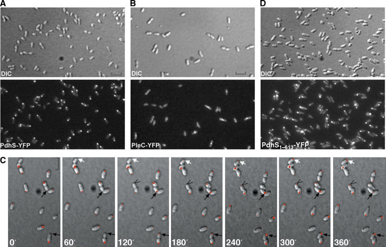
PdhS is dynamically localized at one pole of B. abortus through its N-terminal domain, whereas PleC localizes at mid-cell position. (A) PdhS localizes at one pole of B. abortus. DIC and corresponding fluorescence images were taken of B. abortus cells producing PdhS-YFP (XDB1104 strain). (B) BaPleC localizes at mid-cell in B. abortus. DIC and corresponding fluorescence images were taken of B. abortus cells producing PleC-YFP (XDB1105 strain). (C) PdhS dynamically localizes at the old pole of B. abortus cells. A time-lapse microscopy experiment was performed on the XDB1104 strain expressing pdhS-yfp by taking DIC and corresponding fluorescence images each 60 min. Arrows show that polar fluorescence signal appears 60–120 min following the physical separation of the daughter cells. (D) The N-terminal domain of PdhS is important for its polar localization. DIC and corresponding fluorescence images were taken of B. abortus cells producing PdhS1–613-YFP from the low-copy plasmid pRH407. Scale bar, 2 μm.