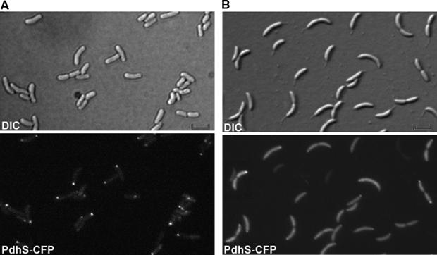
Figure 7.

PdhS localizes at one pole of S. meliloti cells and at the stalked pole of C. crescentus cells. DIC and corresponding fluorescence images were taken with S. meliloti (A) or C. crescentus (B) cells producing PdhS-CFP from the low-copy plasmid pRH324. Scale bar, 2 μm.
