
Figure 7.
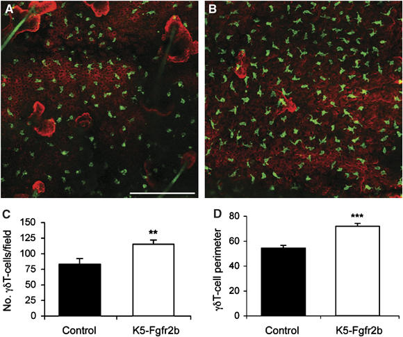
γδT-cell abnormalities in K5-R2b-null epidermis. Whole-mount preparations of ear epidermis from control (A) and K5-R2b-null (B) mice stained with antibodies to γδT cells (green) and keratin 14 (red) highlight the increased γδT-cell density in mutant epidermis (quantitated in (C)). In addition to being present in increased numbers, γδT cells in mutant epidermis display an altered morphology, adopting a more dendritic appearance, reflected by a significantly increased cell perimeter (quantitated in (D)). Scale bar in (A), 50 μm. **P<0.01; ***P<0.001 using the Mann--Whitney test.