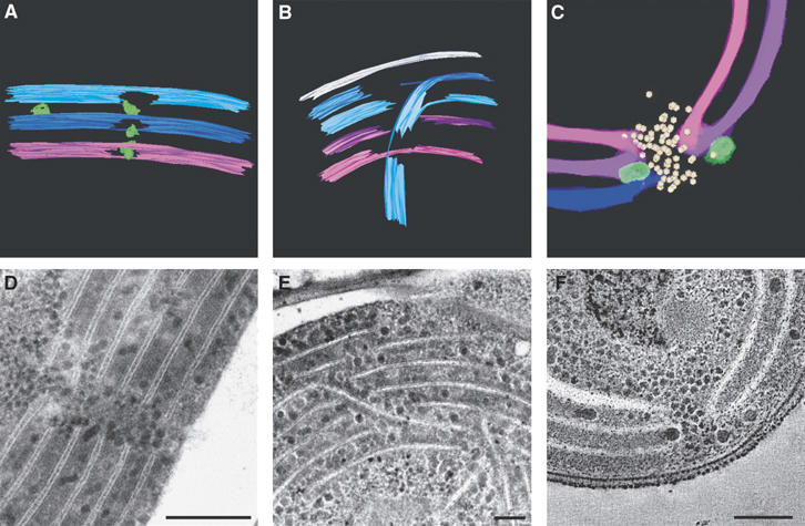
Figure 2.

Types of perforations in the thylakoid membranes. The major types of perforations observed in the thylakoid membranes of Synechococcus sp PCC 7942 and Microcoleus sp are shown. Models (A) generated from a tomogram of a Microcoleus sp cell (not shown), (B) generated from Supplementary Movie S2; (C) an enlargement of the lower perforation seen in Figure 1C; thin-section micrographs of Microcoleus sp cells (D, E), and a tomographic slice (∼12 nm; F, an enlargement of Figure 1A). Scale bars, 200 nm (D); 80 nm (E); 100 nm (F).
