Figure 3.

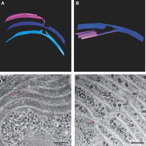
Thylakoid membranes are interconnected. Models (A) generated from a tomogram of a Synechococcus cell (not shown), (B) generated from a tomogram of a Microcoleus cell (see Supplementary Movie S2); and thin-section micrographs of Microcoleus sp cells (C, D), showing how different layers of the thylakoid membranes are connected to each other. Connections involve splitting or branching of the membranes (red arrowheads) and subsequent folding or fusion. A similar mode of connectivity, involving bifurcations and fusion of the membranes, has also been observed in the thylakoid membranes of higher-plant chloroplasts (Shimoni et al, 2005). Scale bars, 100 nm.
