
Figure 8.
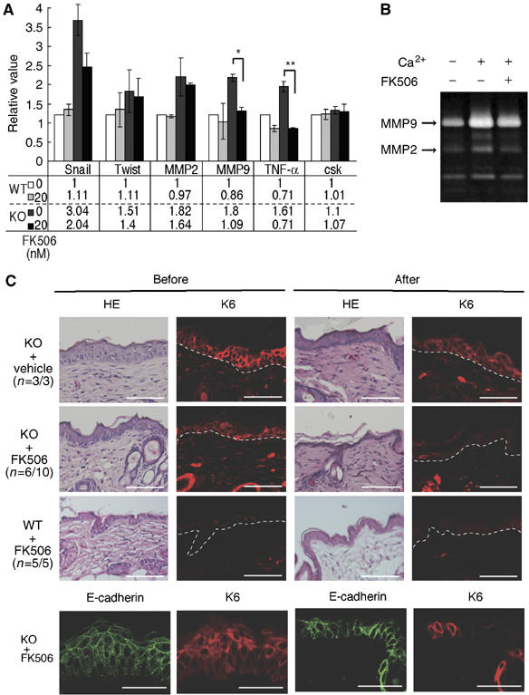
Effects of FK506 treatment on keratinocytes and epidermis in K5 csk-KO mice. (A) Expression of proinflammatory factors and EMT markers in K5 csk-KO keratinocytes. Expression of the indicated genes in wild-type (WT) and K5 csk-KO (KO) keratinocytes treated in the presence or absence of FK506 (20 nM) was quantified by real-time PCR. Values relative to nontreated WT controls are shown in the upper graph (mean±s.e. of five independent experiments, t-test; **P<0.01, *P<0.05). Actual mean values are shown in the lower table. (B) Zymogram of MMP activity. Conditioned medium was prepared from K5 csk-KO keratinocytes treated with the indicated reagents and then subjected to zymography in a gelatin-containing gel. Positions corresponding to MMP9 and MMP2 are indicated by the arrows. (C) The skin of K5 csk-KO mice (KO) was treated with FK506 ointment or control vehicle (petroleum jelly). The skin of WT mice was treated with FK506 as a control. Dorsal skin sections stained with HE and anti-K6 before (left) and after (right) treatment are shown. Frequencies (n=affected individuals/total individuals) are shown in parentheses. The skin of K5 csk-KO mice (KO) was further subjected to staining for E-cadherin before and after treatment with FK506 (lower panels). The dotted white line represents the epidermal–dermal border. Scale bars: 10 μm.