
Figure 6.
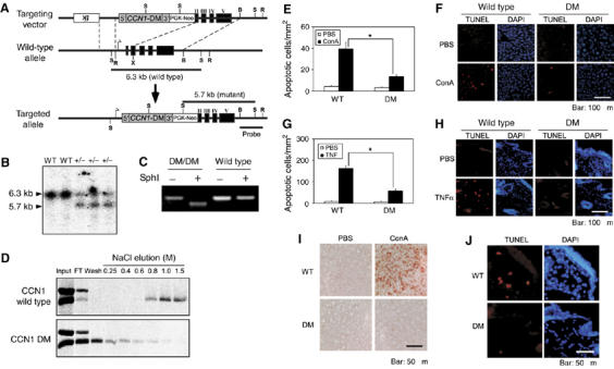
Generation of Ccn1dm/dm mice and blunted TNFα-mediated apoptosis in vivo. (A) A gene targeting construct replaced the EcoRI–XmaI fragment of Ccn1 with a cDNA encoding Ccn1dm. The recombined allele maintained the Ccn1 promoter and transcription start site and expresses the Ccn1dm cDNA that preserved the 5′ and 3′ untranslated sequences. Thymidine kinase (tk) and PGK-neo were used as selectable markers in homologous recombination in ES cells. B, BamHI; X, XmaI; R, EcoRI; S, SphI. (B) DNA samples isolated from wild-type (WT) or Ccn1dm/Ccn1 mice (+/−) were digested with SphI and probed with a BamHI/EcoRI fragment, yielding 6.3 (wild-type) and 5.7 (targeted) kb bands illustrated in (A). (C) Total RNA was isolated from MEFs, reverse-transcribed and the Ccn1 sequence amplified by PCR. The Ccn1dm cDNA contains an engineered diagnostic Sph1 site, which is absent in the wild-type sequence. (D) MEFs from Ccn1dm/dm mice and their wild-type littermates were serum-stimulated to induce synthesis of CCN1 while being labeled with 35S-cysteine. Total cell lysates were passed through a heparin-sepharose column, and eluted with buffer containing varying concentrations of NaCl as indicated. Eluted proteins were immunoprecipitated with anti-CCN1 antibodies, resolved on SDS–PAGE and exposed to X-ray film. (E) ConA (20 mg/kg body weight) was delivered by tail-vein injection, and liver apoptosis analyzed 8 h thereafter by TUNEL assay (labeled with rhodamine). Numbers of apoptotic cells were counted in three randomly chosen fields and expressed as cells/1 mm2 of tissue. (WT, n=5; Ccn1dm/dm, n=8; *P<0.001). Histological sections are shown in (F). (G) Apoptosis was induced by subcutaneous injection of TNFα (400 ng in 50 μl). After 8 h, skin tissue from injection sites was collected, processed, and subjected to TUNEL assay. Numbers of apoptotic cells were counted as above. (WT, n=4; Ccn1dm/dm, n=7; *P<0.001). Histological sections are shown in (H), with a higher magnification view shown in (J). (I) Liver tissue of ConA or PBS-treated WT or Ccn1dm/dm mice were stained with anti-8-OHdG antibodies.