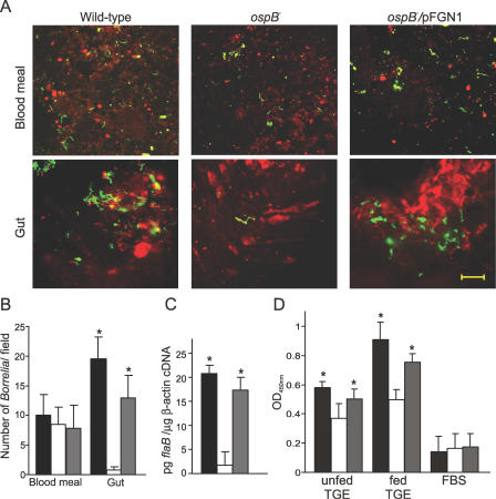
Figure 4. OspB Complementation Restores B. burgdorferi Ability to Survive and Colonize Ticks.
Results from the experiments with the ticks inoculated by microinjection via rectal aperture are shown. Approximately 103 spirochetes were microinjected into each tick.
(A) Shown are representative confocal microscopic images of the blood meal and gut samples of microinjected nymphs at 48 h during feeding. Samples were stained with FITC-labeled anti-Borrelia antibody (green), and tick cell nucleic acid was stained with propidium iodide (red). Scale 20 μM.
(B) Average value of number of spirochetes from 15 random microscopic field observations is shown.
(C) Q-RT-PCR for the total RNA extracted from microinjected nymphs collected at 48 h during feeding is shown. Values on Y-axis represent pg flaB/μg tick β-actin cDNA.
(D) Shown are the readings from an in vitro binding assay of the wild-type, the ospB mutant, and the OspB complemented mutant B. burgdorferi to TGE- and FBS-coated wells (see Materials and Methods for details). Values on Y-axis are sample absorbance measured at 450 nm of wavelength. The values shown are the averages of four independent experiments. Black, open, and gray bars in (B, C, and D) indicate values for the wild-type, the ospB mutant, and the OspB complemented mutant spirochetes, respectively. Error bars define standard deviation (+) from the average value (mean). * indicates values that are statistically significant (p < 0.05, Student t test) in comparison to the ospB mutant.

