
FIG. 5.
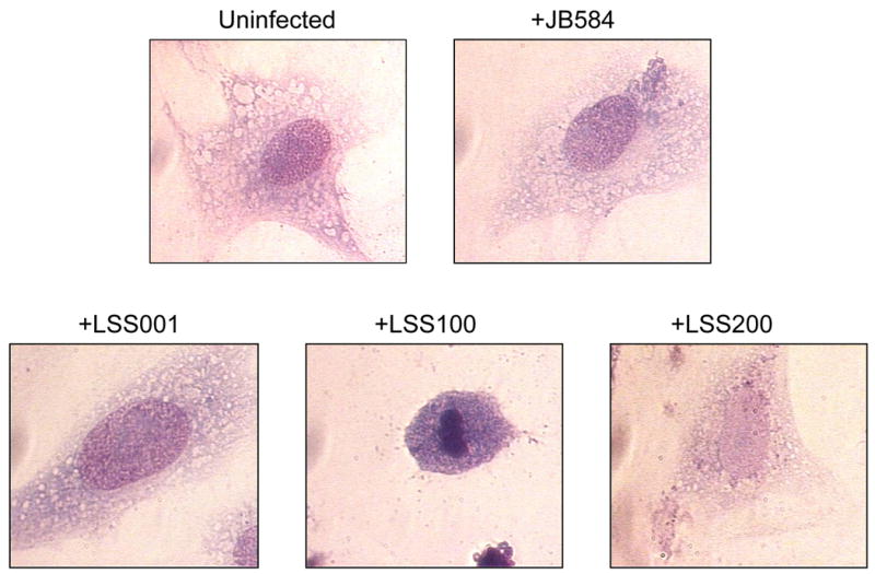
Micrographs showing representative cell morphologies of infected HUVECs following a 96-h co-culture with four B. bacilliformis strains including JB584, LSS001, LSS100 and LSS200. An uninfected HUVEC is provided for comparison. Cells were visualized using Hema-Quik Stain (Fisher Scientific) and are shown at 1000x magnification. Note the marked apoptosis in +LSS100 cells.