
Figure 1. BDNF persistently accelerates the initial phase of evoked release from presynaptic terminals on dendritic spines of eGFP-transfected cultured hippocampal neurons.
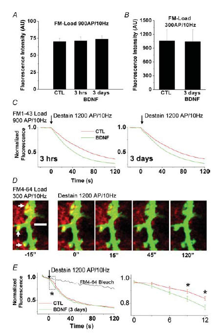
A, initial raw fluorescent intensities of hippocampal presynaptic terminals from control and brain-derived neurotrophic factor (BDNF)-treated cultures (3 h and 3 days) following FM1-43 staining with a strong stimulus (900 APs at 10 Hz) loading ∼80% of the total recycling pool, as estimated by high K+ stimulation (Student's t test, P > 0.05). B, initial raw fluorescent intensities of hippocampal excitatory presynaptic terminals on dendritic spines of eGFP-transfected control and BDNF-treated neurons (3 days) following FM4-64 staining with a weaker stimulus (300 APs at 10 Hz) loading ∼40% of the total recycling pool, as estimated by high K+ stimulation (Student's t test, P > 0.05). C, either 3 h or 3 days exposure to BDNF accelerated FM1-43 destaining evoked by field electrical stimulation (1200 APs at 10 Hz). D, time-lapse confocal images of individual hippocampal excitatory presynaptic nerve terminals (FM4-64, red puncta) overlapping onto dendritic spines of a control postsynaptic neuron (eGFP, green). Time-lapse images were acquired for baseline, as well as during the delivery of the field stimulation protocol used to evoke synaptic vesicle exocytosis. Note the decrease in brightness of red puncta across time (left to right) corresponding to a decrease in FM4-64 fluorescence as a result of exocytosis. E, background-subtracted line plots of mean normalized pixel intensities during baseline acquisition, and during field stimulation, for terminals from control (red) and BDNF-treated cultures (green). The area in the box is enlarged in the plot to the right to highlight the effect of BDNF on FM destaining during the initial phase of the stimulation protocol. Asterisks indicate significant differences (ANOVA followed by Scheffé's procedure for multiple comparisons, P < 0.05).