
Figure 1. 5-Hydroxyindole potentiates currents through the rat native α7 nAChR.
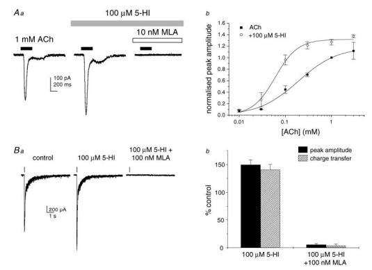
Aa, representative ACh-evoked inward currents in rat hippocampal neurons in primary culture. Currents were enhanced in the presence of 5-hydroxyindole (5-HI; 100 μm) and abolished by methyllycaconitine (MLA; 10 nm). Ab, concentration–response curves for ACh in the absence (▪) and in the presence (○) of 5-HI (100 μm; n = 4 cells per data point). In the presence of 5-HI, the ACh EC50 shifted from 194 to 65 μm (P < 0.05) and the Hill slope increased from 1.2 to 2.1 (P < 0.05). The maximal efficacy was not significantly affected. Ba, representative fast inward currents elicited by pressure microejection of ACh (1 mm) onto CA1 stratum radiatum interneurons in a rat hippocampal slice. The responses were observed in the presence of picrotoxin (100 μm), 2,3-dioxo-6-nitro-1,2,3,4-tetrahydrobenzo(f)quinoxaline-7-sulphonamide disodium (NBQX; 5 μm), d-aminophosphonovalerate (d-AP5; 50 μm), MDL72222 (0.5 μm) and dihydro-β-erythroidine (DHβE; 0.1 μm). The time course of agonist application is indicated by the bars above the traces. Bb, mean agonist-evoked currents were enhanced in the presence of 5-HI (100 μμ) and inhibited in the presence of MLA (100 nm; n = 5–8 cells).