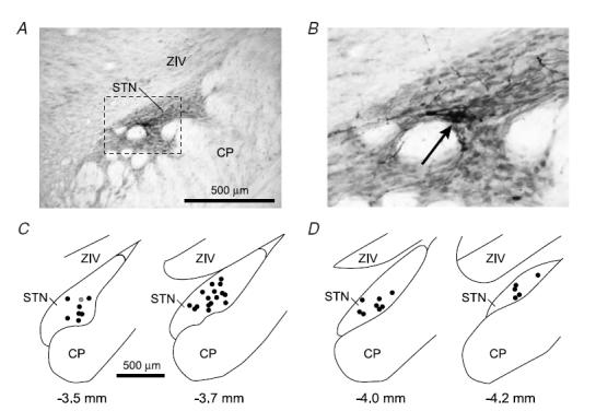
Figure 1. Locations of recorded subthalamic nucleus neurons.

A, digital micrograph of a typical Neurobiotin deposit that marked a recording location in the rostral half of the subthalamic nucleus (STN). Tissue section cut in the coronal plane (dorsal towards the top, lateral to the right). The STN is characterized by a higher density of neurons compared to the overlying ventral division of the zona incerta (ZIV). CP, cerebral peduncle. b, area in A within the dashed box shown at greater resolution. The core of the Neurobiotin deposit is identified as a dense accumulation of black reaction product (arrow), which is accompanied by Golgi-like labelling of neighbouring neurons. C, locations of all 24 neurons recorded in the rostral half of the STN, plotted in the coronal plane. The number below each section corresponds to the distance posterior of bregma. Location shown in A is highlighted as a grey circle. D, locations of all 13 neurons recorded in the caudal half of STN. Scale bar in C also applies to D.
