
Figure 4.
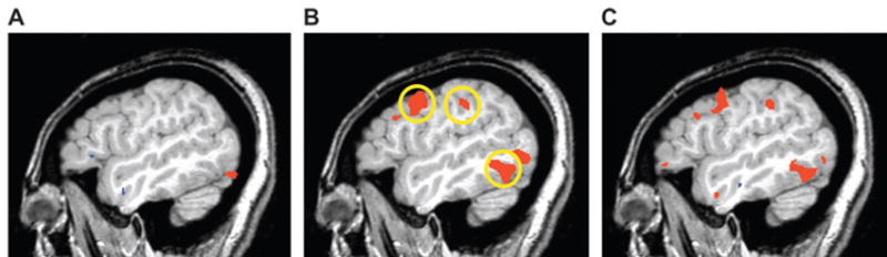
Hemodynamic response in a single subject prior to and after training. Shown is a single, 1-mm-thick magnetic resonance image of the left hemisphere (x = −47). Regions in red were significantly more active when performing the matching task with the novel objects relative to scrambled object matching. (A) Prior to training, the object-matching task was associated with activity in ventral occipital cortex, which continued anteriorly along the fusiform gyrus (not visible in this lateral slice). (B) After training, activity emerged in the left middle temporal gyrus and left premotor and intraparietal cortices (circled regions). (C) Direct comparison of the pre- and posttraining scans revealed significantly more activity in these regions after training (all P < 0.001).