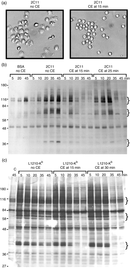
Figure 3.
Cytochalasin E (CE) disrupts ongoing tyrosine phosphorylation stimulated with immobilized anti-CD3 or target cells. (a) Cl 11 cells were added to wells pre-coated with 145-2C11 (anti-CD3). After 15 min on the plates, carrier (left panel) or CE to a final concentration of 10 µm (right panel) was added to the cells. Photographs of the cells were taken within 5 min after addition of the inhibitor. Cl 11 cells were stimulated for the indicated time with (b) bovine serum albumin (BSA) or immobilized anti-CD3 or (c) L1210 target cells transfected with Kb. In both cases, CE was added to Cl 11 at a final concentration of 10 µm at the indicated time after addition of cells to immobilized 145-2C11 (b) or mixing with target cells (c). At the indicated time, cells were lysed with reducing sample buffer, and subjected to sodium dodecyl sulphate–polyacrylamide gel electrophoresis (SDS-PAGE) and immunoblotting with antiphosphotyrosine antibodies. Lane ‘C’ in (c) refers to Cl 11 cells mixed with untransfected L1210 cells. The curly brackets indicate positions of groups of proteins that become dephosphorylated after CE addition. Molecular weight standards (kDa) are shown to the left of the gels in (b) and (c). The data shown are representative of four different experiments with Cl 11 or AB.1.

