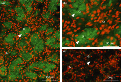
Figure 7.
Immunohistochemical staining for interleukin (IL)-1β in the submandibular gland (SMG) of lipopolysaccharide (LPS)-injected C3H/HeN mice. The SMG was removed 6 hr after LPS injection. (a, c) Low-power magnification. (b) High-power magnification. (a, b) Staining with anti-IL-1β antibody immunoglobulin (IgG). (c) Staining with antigen-pre-absorbed antibody IgG. Arrows indicate secretory granules of granular convoluted tubule (GCT) cells. Red staining indicates the location of the nucleus, whereas green staining indicates the location of IL-1β. The GCT cells (G) filled with immunopositive secretory granules have nuclei in the basal area. No positive reaction was seen in acini (Ac).

