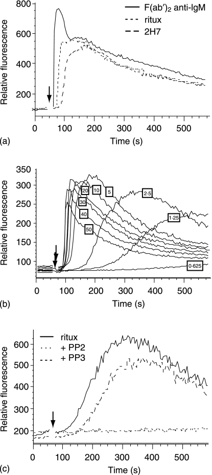
Figure 2.
Rituximab hypercrosslinking induces SFK-dependent calcium mobilization. (a) Ramos B cells were loaded with fluo-3AM and then incubated with rituximab or 2H7. After establishing the baseline, samples were stimulated with 50 µg GAH or GAM (arrow). Unligated cell samples were stimulated with anti-IgM for comparison (solid line). (b) Cells incubated with rituximab were stimulated with concentrations of GAH ranging from 0·625 to 50 µg, as indicated. (c) Cells were untreated or treated with 10 µm PP2 or PP3, then incubated with rituximab. After establishing the baseline, cells were stimulated with 5 µg of GAH. Data are representative of at least three independent experiments. The scales on the y-axes show arbitrary units assigned by the FlowJo analysis program and are only comparable for samples run in the same experiment.

