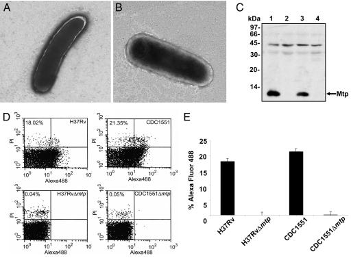
Fig. 2.
Isogenic M. tuberculosis mtp mutants lack production of pili fibers. (A and B) Electron micrographs of H37RvΔ mtp (A) and CDC1551Δ mtp (B) showing no filamentous structures. (Magnification: ×25,000.) (C) Anti-Mtp Western blot of whole bacteria extracts showing production of the pilus protein in H37Rv (lane 1) and CDC1551 (lane 3) and absence of the pilin in both H37RvΔ mtp (lane 2) and CDC1551Δ mtp (lane 4). (D) Surface detection of MTP in wild-type parental and derivative mtp deletion strains by flow cytometry (n = 50,000) using anti-Mtp antisera. MTP–antibody complexes were detected by using Alexa Fluor conjugate. (E) Plot of the flow cytometry values obtained in D.

