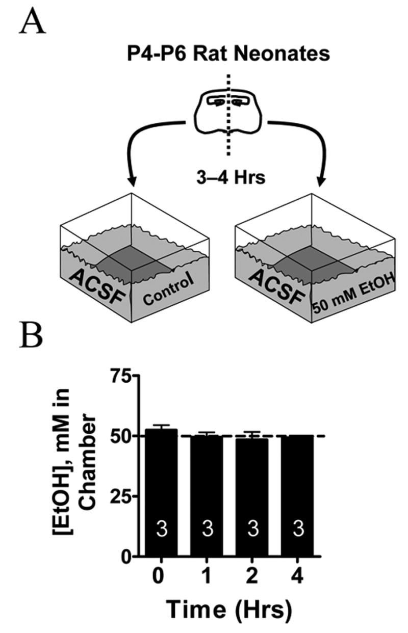
Fig. 1. In Vitro EtOH Exposure Paradigm.

(A) Scheme illustrating the procedure used to expose neonatal (P4–P6) hippocampal slices to 50 mM EtOH for 3–4 hrs. Sister slices were segregated into a chamber that contained continuously oxygenated ACSF solution with or without EtOH. Left and right sister slices were randomly assigned to each chamber. All slices were allowed to equilibrate for a period greater than 80 min prior to the start of the experiment. (B) EtOH concentrations in the ACSF do not significantly change as a function of time. Numbers in each bar represent the number of experiments.
