
Fig. 2.
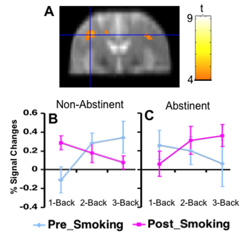
A cluster in the left dorsal lateral prefrontal cortex (DLPFC) showed a 3-way interaction among acute smoking, test session, and task load on task-related activity. Colors superimposed on the gray scale image, from the study-specific structural brain template, indicate values of t according to the color bar. The line graph indicates the mean percent signal changes from the 0-back and standard deviation at each level of the N-Back Task. B. data from session after smoking ad libitum, C: data from session after overnight abstinence.