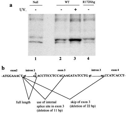
Figure 2.
(a) Analysis of p53 protein levels in fibroblasts from embryos null for p53 (lane 1), wild-type (WT; lanes 2 and 3), or homozygous for the p53R172HΔg mutation (lane 4). Wild-type fibroblasts were treated with UV to show induction of p53 and confirm position on the gel (lane 3). (b) The structure of the p53 gene around exons 2–4 and the spliced products identified in fibroblasts from homozygous p53R172HΔg mutant mice. Δ denotes the G nucleotide in intron 2 that is deleted.

