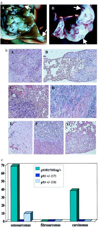
Figure 5.
Frequency of metastasis from tumors of p53R172HΔg/+ mice. (a) Metastatic lesions from an osteosarcoma that developed in a p53R172HΔg/+ mouse. (A) Liver. (B) Lung. (b) Representative histologies of tumors and their metastatic spread. A, C, and E represent primary tumors; B, D, F, and G represent their corresponding metastases. (A and B) Hepatocelluar carcinomas with metastasis in lung. (C and D) Lung adenocarcinomas with metastasis in myocardium (E, F, and G) Osteosarcoma with metastasis in liver (F) and lung (G). (c) Frequency of metastasis from p53R172HΔg/+ mice vs. p53+/− mice. p53+/− metastasis data are from Harvey et al. (17) and Tervana et al. (18), respectively.

