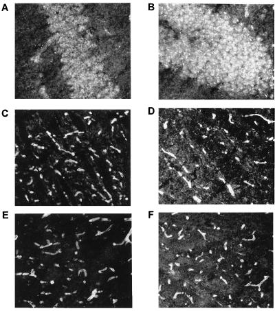
Figure 5.
Immunolocalization of β-dystrobrevin in the brain. Coronal mouse brain sections were stained with the following antibodies: anti-β-dystrobrevin, β521 (A and B), anti-α-dystrobrevin 1, α1CT-FP (C), anti-dystrobrevin, ab308 (D), agrin IIA (E), and anti-dystrophin 1538 (F). β521 stains neurons in the pyramidal cell layer of the hippocampus and the dentate gyrus (A and B). The other antibodies label the microvasculature. Magnification: ×400 (A and B) and ×200 (C-F).

