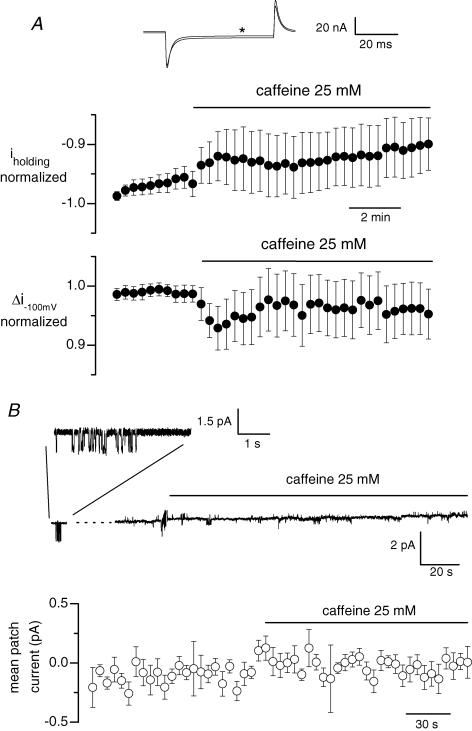
Figure 8. Effect of caffeine on whole-cell background current and single channel activity in fibres that were free of silicone.
In A, fibres were voltage-clamped using the whole-cell configuration of the patch-clamp technique with the pipette solution containing 20 mm internal EGTA. The holding voltage was set to −80 mV and 50 ms pulses to −100 mV were applied every 20 s. The upper panel shows the last membrane current record measured in response to a 20 mV hyperpolarization in the presence of the control external solution, superimposed on the membrane current record obtained in response to the same pulse applied 8 min and 40 s after addition of caffeine (record marked by a star). The middle and lower panels show the mean normalized value of the whole-cell background current at −80 mV and the mean change in membrane current induced by the 20 mV hyperpolarization, respectively, along the course of such experiments. Data are from 12 cells. In B, the upper panel shows the single channel activity recorded in a cell-attached patch established on a fibre previously bathed for 30 min in a Tyrode solution containing 250 μm EGTA-AM. The holding pipette potential was 0 mV. Activity of channels carrying inward current, illustrated on an expanded scale in the inset shown on top, was detected 2 min 30 s before the beginning of the continuous recording. The lower panel shows mean values for the patch current from 8 identical experiments. In each single channel record, the closed channel state was set to zero and the current value was averaged over successive intervals of 5 s; the graph shows the corresponding mean current versus time.

