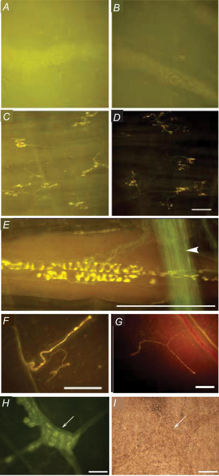
Figure 8. Stimulation- and muscarinic receptor-independent labelling of neuronal structures by the styryl dye FM2-10.
Representative photomicrographs showing FM2-10 labelling of the guinea-pig myenteric plexus (A and B) and mucosal afferent nerve terminals in the trachea (C and D) in the absence (A and C) and presence (B and D) of 1 μm atropine. Note that atropine substantially reduces the background fluorescence in both tissues without preventing staining of the nerves in either the myenteric plexus or the airway mucosa. E, intravital labelling of a sensory nerve terminal associated with the abdominal muscles in guinea-pigs. Labelling was carried out in the presence of atropine and occurred without any stimulation of the tissue. Also note the large nerve bundle stained green in the foreground of the image (arrowhead). Less complex neuronal structures were similarly labelled by FM2-10 in the presence of atropine in the guinea-pig (F) and rat membranous diaphragm (G). These structures did not appear to be motor nerve terminals and are presumably diaphragm stretch receptors. As with all structures labelled in this figure, labelling occurred independent of any overt attempt to stimulate these nerves. H, parasympathetic ganglia associated with the rat trachea were also labelled by FM2-10 in the presence of atropine; the same results were also seen with guinea-pig trachea (not shown). I, shows the ganglia in H when viewed in bright field. Scale bar, 50 μm (A–D, H and I) and 100 μm (E–G).

