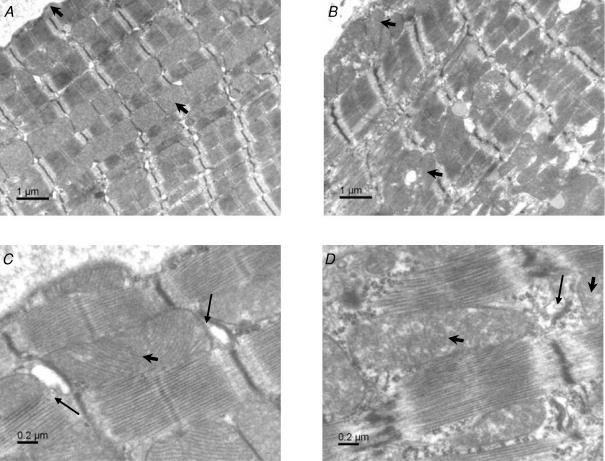
Figure 1. Electron microscopic images of left ventricle from control and MLP-null mice.
A, overview of a control myocyte in longitudinal section, with mitochondria arranged in longitudinal columns. B, overview of an MLP-null cardiomyocyte, showing myofibrillar disorganization, an irregular arrangement of intermyofibrillar mitochondria, and increased content of subsarcolemmal mitochondria. C, detail of sarcomeres in a control myocyte, showing mitochondria tightly packed with SR. D, detail of sarcomeres in an MLP-null myocyte, showing looser packing of mitochondria with SR, irregular and widened Z-lines, and cytoplasm in the intermyofibrillar space. Arrowheads show mitochondria, arrows show SR.

