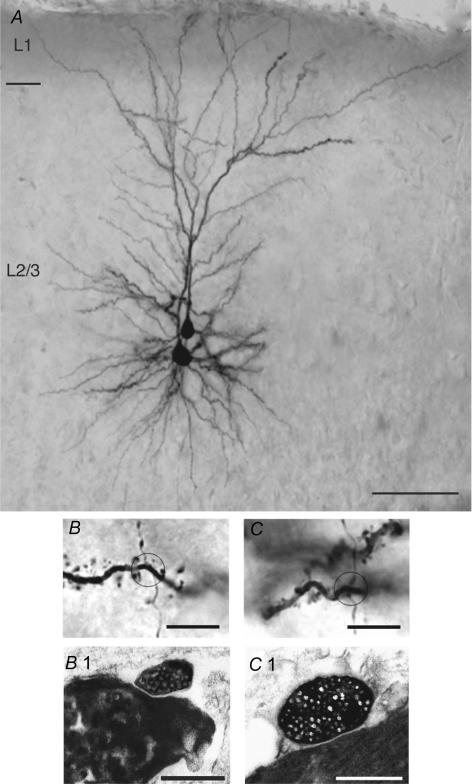
Figure 4. Half-tone image of a pair of synaptically coupled L2/3 pyramidal cells including the light and electron microscopic identification of synaptic contacts.
A, low magnification light microscopic image of two synaptically coupled pyramidal cells filled with biocytin. Both pyramidal cells were located in the middle portion of layer 2/3. Note the elaborate symmetric basal dendritic field and the apical dendrites forming extensive tufts terminating in layer 1. Calibration bar, 100 μm. B and C, high magnification of the synaptic contacts established by en passant axonal collaterals of the presynaptic neurone on different basal dendrites of the postsynaptic L2/3 pyramidal cells. The calibration bar is 5.0 μm for both panels. B1 and C1, both light microscopically identified synaptic contacts were identified at the electron microscopic level. The synaptic boutons of the presynaptic axon collaterals are clearly identifiable by their content of transmitter vesicles. The calibration bar is 1.0 μm for both panels.

