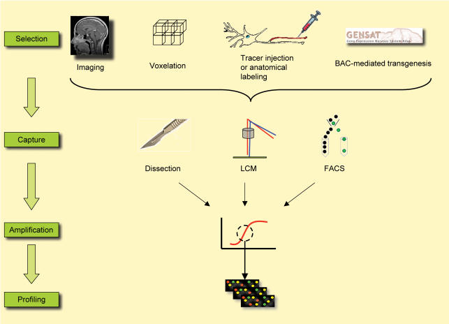
Figure 1. Schematic illustrating the major methods currently available to address regional and cell specificity in human brain and experimental models.
In human brain, neuroanatomical methods can be used to guide dissection and laser-guided microdissection. Voxelation approaches allow visualization of 3D gene expression maps. In experimental models, in addition to image-based methods, cell specificity can be achieved with labelling methods, based on both neuroanatomical and genetic markers (e.g. http://www.ncbi.nlm.nih.gov/projects/gensat/). Such intrinsic labels can be used to guide dissection or FACS-based approaches. Most of these methods require an amplification step due to low RNA amounts, prior to gene expression profiling using microarrays. Many robust methods for such amplification are now widely available, making this a worthwhile approach.

