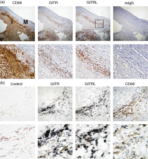
Figure 1.
Macrophages express GITR and GITRL in the atherosclerotic plaques. (a) Plaque sections were stained with antibodies against CD68, GITR, or GITRL. Isotype-matching control antibody (mouse IgG1) was used to demonstrate the specificity of the staining. Low magnification (× 100) pictures of consecutive sections of atherosclerotic plaque are shown in the upper panel and high magnification (× 400) pictures are shown in the lower panel. I, intima; M, media. (b) Serial sections of an atherosclerotic plaque were used for in situ hybridization using GITR- or GITRL-specific probes or a non-specific control probe as described in the Materials and methods section. The hybridization was visualized as dark purple. For comparison, an adjacent section was immunostained using anti-CD68 mAb (brown colour). Magnifications are × 400 for the upper panel and × 1000 for the lower panel.

