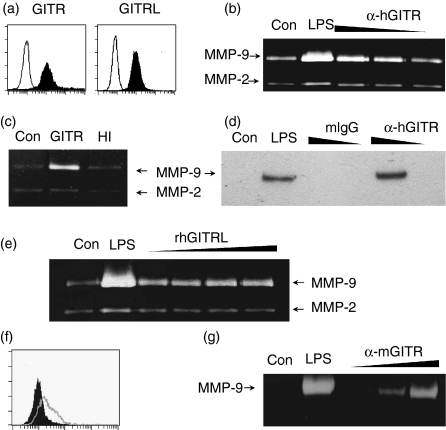
Figure 2.
Stimulation of GITR induces MMP-9 expression in monocyte/macrophage cell lines. (a) THP-1 cells were stained with anti-GITR or anti-GITRL mAbs as indicated. Fluorescence profiles obtained from specific staining (filled area) and background staining (open area, stained with isotype-matching control antibody) are compared. (b) THP-1 cells were stimulated with anti-hGITR mAb immobilized at 20, 6 and 2 μg/ml concentrations. Culture supernatants were collected over a 24 hr period to measure MMP-9 activity using gelatin zymogram. (c) THP-1 cells were stimulated with immobilized anti-GITR mAb (10 μg/ml) that had been pretreated with (HI) or without (GITR) heat (95° for 30 min). (d) THP-1 cells were stimulated with anti-GITR mAb or mIgG immobilized at 20 and 2 μg/ml concentrations. Culture supernatants were collected after 24 hr and condensed approximately 10-fold before Western blot analysis with anti-MMP-9 mAb. (e) THP-1 cells were stimulated with 0·1, 0·3, 1 and 3 μg/ml of rhGITRL for 24 hr before gelatin zymogram. (f) RAW 264.7 cells were stained with anti-GITR mAb. Histograms from specific staining (open area) and background staining (filled area, stained with isotype-matching control antibody) are compared. (g) RAW264.7 cells were stimulated with anti-mGITR mAb which was added to culture medium at 1, 10 and 30 μg/ml concentrations. Culture supernatants were collected after 48 hr for gelatin zymogram. As a positive control, the cells were treated with 1 μg/ml lipopolysaccharide (LPS). Con, no treatment control.

