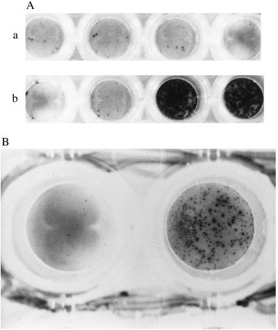
Figure 2.
(A) Photomicrograph showing peptide-specific HLA-B52-restricted IFN-γ release by ES12-specific CD8+ clones derived from donor NPH54. Cells from clones 3–1, 3–15, and 3–98 were pooled and assayed against unpulsed (first pair of wells in a and b), and ES12-pulsed (second pair of wells in a and b). HLA-B52-mismatched (A) and matched (B) target BCLs. The mismatched BCL is PG: HLA-A2.01, HLA-A3; HLA-B7, HLA-B51 and is shown in A. The matched BCL, a homozygous typing line, is Akiba: HLA-A24; HLA-B52, and is shown in B. Assays were performed in duplicate wells with 5,000 T cells and 50,000 B cells per well. Only the pair of duplicate wells with ES12-pulsed HLA-B52-matched targets are positive; the spots are so numerous that they appear confluent. (B) Photomicrograph showing IFN-γ release by ES12-specific CD8+ clone 3–15 of donor NPH54, in response to autologous BCL infected with vaccinia virus recombinant for the gene encoding ESAT-6 (rVV-ESAT-6). The negative control is the left-hand well, using autologous BCL infected with rVV control. BCL were infected the night before with the respective recombinant viruses at a multiplicity of infection (m.o.i) of 7 plaque-forming units per cell in serum-free medium; after 90 min, cells were diluted up to 1 million/ml in R10 and incubated overnight. Infected BCL (100,000) were then added to each well along with 5,000 cloned T cells. The photomicrograph shows the result with clone 3–15, giving in excess of 450 SFCs. The results with the other two clones, 3–1 and 3–98, were so strongly positive that the spots were confluent. (×20.)

