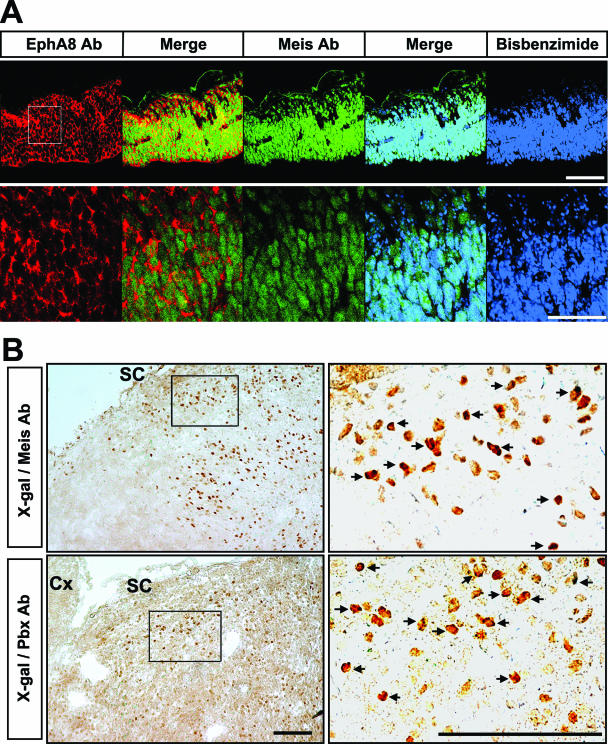
FIG. 6.
Colocalization of Meis, Pbx, and EphA8 proteins in the anterior region of the mesencephalon. (A, top) Immunohistochemical analyses of EphA8 (red) and Meis (green) proteins in the dorsal mesencephalic tissues at E11.5. Scale bar, 100 μm. (Bottom) Enlarged views of each image shown in panel A (the area of the enlargement is boxed in the left-hand upper image). Scale bar, 50 μm. (B) X-Gal staining of sagittal sections of the superior colliculus of P0 ephA8+/lacZ mouse, followed by incubation with Meis (top row) or Pbx (bottom row) antibody. The right-hand images are enlarged views of each box shown in the left-hand images. The arrows mark a subset of cells coexpressing either Meis or Pbx with the EphA8 receptor. SC, superior colliculus; Cx, cortex. Scale bar, 10 μm.

