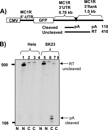
FIG. 1.
The MC1R reporter gene shows cell type-specific expression patterns. A) Diagram depicting the MC1R reporter gene. The position and the lengths of the MC1R-derived UTRs are indicated above the diagram. The promoter (CMV) and the GFP open reading frame (GFP) are represented by open boxes. The MC1R poly(A) site is pointed out by a filled triangle and pA. RNase protection fragments representing uncleaved (RT) and cleaved (pA) RNA species are shown as black lines under the diagram, and the expected lengths of the protected bands are given on the right side. B) RNase protection analysis of nuclear (N) and cytoplasmic (C) RNA isolated from HeLa cells (lanes 1 to 4) and melanoma-derived SK23 cells (lanes 5 to 8), which were transiently transfected with the MC1R wt reporter construct. Arrows with pA and RT indicate RNase protection bands representing cleaved and uncleaved MC1R RNA species. Even-numbered lanes are control transfections where the VA plasmid is cotransfected with an empty pUC18 vector to control for potential stimulation of endogenous MC1R expression. Note that RNA was normalized to the cotransfectional VA control.

