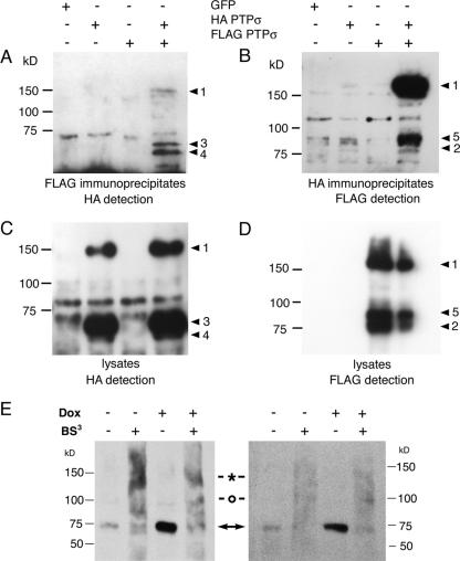
FIG. 3.
Wild-type PTPσ homodimer detection. (A to D) Tagged PTPσ proteins were transfected into 293T cells and immunoprecipitated with either anti-FLAG (A) or anti-HA (B). The immunoblots were then detected with either anti-HA (A) or anti-FLAG (B), respectively. The corresponding lysates are shown in panels C and D. The PTPσ protein band numbers to the right of each panel correspond to those described in the legend to Fig. 2E. Panel E shows PTPσ proteins expressed under doxycycline-inducible control in a PC12-derived cell line, 12CRYP11, detected with IG2 serum (52). Two experimental examples are shown in which cells were treated with or without doxycycline (Dox) and with or without the extracellular cross-linker BS3. The native 75-kDa proteins are indicated with arrows, and commonly seen cross-linked 140-kDa and 100-kDa bands are indicated with an asterisk and a circle, respectively. The gels were run under nonreducing conditions because the epitope(s) detected by IG2 antibody is redox sensitive.

