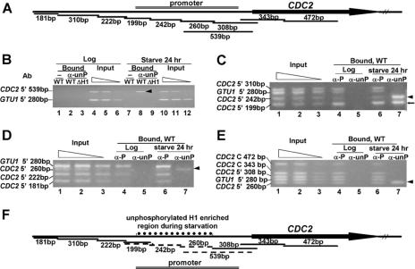
FIG. 3.
Unphosphorylated H1 is specifically localized to a restricted region of the CDC2 promoter during starvation when CDC2 is weakly expressed. A. Diagram of the CDC2 PCR fragments analyzed in the ChIP assay. Primer sets that amplify adjacent fragments differing in length by 19 to 130 bp were designed across the 5′ and coding regions of the CDC2 gene. The 539-bp fragment overlaps two fragments (260 bp and 308 bp) that were also analyzed. Different fragments were amplified in multiplex PCR of input or immunoprecipitated (Bound) DNA together with a 280-bp control fragment immediately upstream of the GTU1 coding region. GTU1 gene expression is not affected by H1 phosphorylation. B. Chromatin in the CDC2 promoter is enriched in unphosphorylated H1 in starved cells. ChIP was done on log-phase or 24-h-starved WT CU428 cells or H1 knockout cells (ΔH1) using purified anti-unphosphorylated H1 antibody (α-unP) or no antibody (−). Input DNA and immunoprecipitated DNA were purified and used in multiplex PCR to amplify a 539-bp fragment immediately upstream of the CDC2 coding region and the GTU1 280-bp fragment. The input sample was diluted in a 2.5-fold series to serve as a quantitation control. Shown are the PCR products from the bound or input DNA analyzed on a 3% agarose gel and stained with ethidium bromide. The arrowhead shows the CDC2 539-bp fragment that was enriched by immunoprecipitation with purified anti-unphosphorylated H1 antibody only in starved-cell chromatin. No PCR fragments were detectable in the absence of antibody or in chromatin from cells lacking H1. C, D, and E. Mapping the region enriched in unphosphorylated H1 in starved cells. ChIP was done on log-phase or starved cells, and the immunoprecipitated DNA was analyzed as described above. The input results are for the starved cells, where the critical observations have been obtained. The inputs from the growing cells are indistinguishable from those of starved cells (data not shown). Arrowheads indicate the fragments that were enriched by purified anti-unphosphorylated H1 antibody in starved cells. The arrow (←) in panel C points to a fragment that was enriched by immunoprecipitation with both anti-phosphorylated H1 (α-P) and purified anti-unphosphorylated H1 (α-unP) antibodies. F. Summary of the ChIP results. Fragments reproducibly enriched by immunoprecipitation with anti-unphosphorylated H1 antibody in 24-h-starved cells are indicated by dashed lines. Fragments that were not enriched were labeled by solid lines. The left boundary of the region containing unphosphorylated H1 resides within the 199-bp fragment, since it was enriched by both anti-phosphorylated and anti-unphosphorylated H1 antibodies. The 539-bp fragment contains the right-hand boundary, indicated by the fact that it can be split into one (260-bp) fragment that is enriched in unphosphorylated H1 and another (308-bp) fragment that is not.

