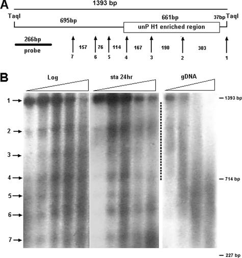
FIG. 4.
Localization of unphosphorylated H1 occurs without major alteration of chromatin structure. Macronuclei from log-phase and 24-h-starved WT CU428 cells as well as genomic DNA from CU428 cells were digested with MNase, followed by TaqI digestion, and analyzed by Southern blotting. A. A diagram of the mapped region. The probe used in panel B is indicated. The numbered arrows correspond to the bands shown in panel B. The numbers in between the arrows are the spacing (in base pairs) between the bands. B. Southern blot showing DNA isolated from macronuclei from log-phase and starved cells as well as genomic DNA. All samples were analyzed on the same gel from which the last, overdigested lane of each sample set and empty lanes separating the different sets of samples were removed to facilitate visual comparisons. The dotted line indicates the position of the part of the blot corresponding to the region enriched in unphosphorylated H1.

