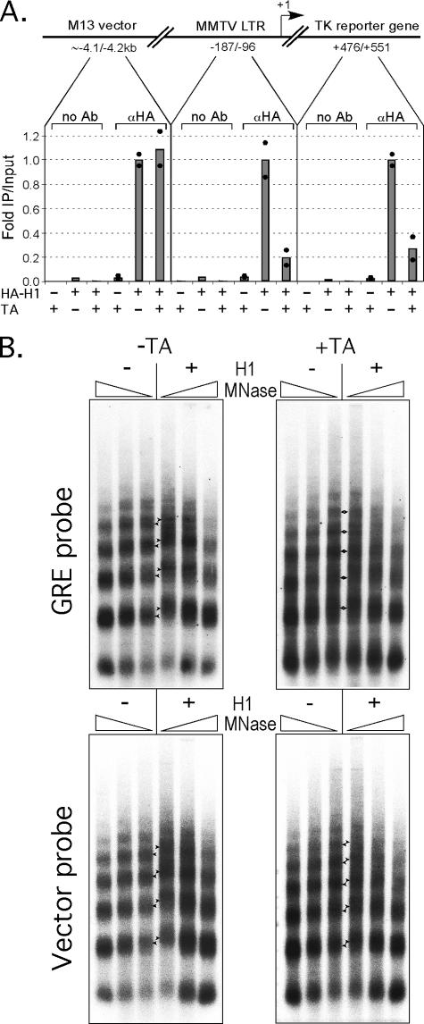
FIG. 7.
(A) Histone H1 dissociates from the promoter and the transcribed region upon hormone activation. Oocytes were injected as described for Fig. 3A and with 0.6 ng of HA-H1 mRNA, followed by ChIP analysis in duplicate. The average amount of DNA relative to input (gray staples) precipitated for samples not treated with hormone was 1.25% at the promoter region, 1.34% at the TK reporter gene, and 0.57% at the vector region. These precipitated amounts were normalized to 1 in the diagram; black dots represent double samples of two independent immunoprecipitations (IPs). (B) Oocytes were injected with GR, NF1, and Oct1 mRNA mix as described for Fig. 3A and also with H1 mRNA to obtain an apparent H1/N ratio of ∼1.3. Hormone treatment (TA) was as indicated: +, present; −, absent. For MNase digestion and analysis, see Fig. 3B. The filter was hybridized with 32P-labeled GRE probe (Fig. 1A) and then, after stripping, rehybridized with 32P-labeled M13 probe.

