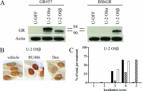
FIG. 4.
Expression and RU-486-dependent nuclear translocation of wild-type hGRβ in the U-2 OSβ stable cell line. (A) The U-2 OS cell line U-2 OSβ, stably expressing hGRβ under the control of a Tet-OFF promoter system, was created. U-2 OSα, U-2 OSβ, and the U-OFF parental cell line were treated for at least 3 h with 1 μM RU-486 and then harvested for Western blot assays. The GR#57 antibody recognizes both hGRα (94 kDa) and hGRβ (90 kDa); the BShGR antibody recognizes hGRβ only; the actin antibody was used to demonstrate equal loading of the lanes. The U-OFF cells did not express detectable levels of GR, while U-2 OSα and U-2 OSβ exclusively expressed hGRα or hGRβ, respectively. Numbers in the middle are molecular masses in kilodaltons. (B) U-2 OSβ cells were treated with ethanol vehicle or 1 μM RU-486 ordexamethasone (Dex) for 3 h before being processed for immunocytochemistry with the GR#57 antibody. Bar, 25 μm. RU-486 but not dexamethasone caused nuclear translocation of stably expressed hGRβ. (C) Quantification of the immunocytochemical localization of hGRβ receptor was carried out by assigning localization scores and plotting a frequency histogram as in Fig. 2B. This analysis confirmed that RU-486 but not dexamethasone caused the wild-type receptor to undergo nuclear translocation. Black bars indicate vehicle treatment; striped bars indicate 1 μM RU-486; white bars indicate 1 μM dexamethasone; n ≥ 290.

