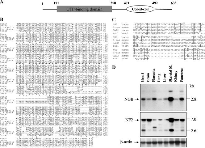
FIG. 1.
Putative domain structure, protein sequence alignment, and expression pattern of NGB. (A) Schematic representation of domain structure of NGB protein, which is composed of a GTP binding domain at the N terminus and a coiled-coil region at the C terminus. (B) Alignment of amino acid sequence of NGB from human, mouse, Drosophila, C. elegans, and yeast cells. Conserved residues are indicated by asterisks. (C) Sequence comparison of GTP-binding domain between NGB, R-Ras, RAP2, and rasl. Conserved amino acids are boxed. (D) Northern blot analysis. A human multiple-tissue mRNA blot was hybridized with [32P]dCTP-labeled NGB, NF2, and β-actin cDNA probes. M., muscle.

