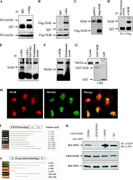
FIG. 2.
Interaction between NF2 and NGB. (A and B) NGB binds to NF2/merlin in HEK293 cells cotransfected with HA-NF2 and Flag-NGB. After 36 h of transfection, cells were lysed, immunoprecipitated with anti-Flag (α-Flag), and detected with anti-HA (α-HA) antibody (A) or vice versa (B). Bottom panels show expression of transfected plasmids. (C and D) Specificity of anti-NGB antibody. HeLa cells were transiently transfected with Flag-NGB, lysed, and then subjected to immunoblotting (C) or IP-Western blotting analysis (D) with indicated antibodies. (E and F) NGB interacts with NF2 at physiological protein levels. 82HTB rhabdomyosarcoma cells were lysed and immunoprecipitated with anti-NF2 (α-NF2) antibody or the antibody was preincubated with GST-NF2 antigen or preimmune serum. The immunoprecipitates were detected with anti-NGB (α-NGB) antibody (E). Conversely, the NGB immunoprecipitates were blotted with anti-NF2 antibody (F). (G) GST-NGB pull down of merlin. A GST pull-down assay was performed by incubation of GST and GST-NGB with HeLa cell lysate. After being washed four times, pull-down products were immunoblotted with anti-NF2 antibody (top). The bottom panel is an SDS-PAGE gel stained with Coomassie blue (CBS). (H) NGB colocalizes with NF2 in the perinuclear region. 82HTB rhabdomyosarcoma cells were cultured on glass coverslips, probed with anti-NGB and -NF2 antibodies, incubated with fluorescein isothiocyanate- and rhodamine-conjugated secondary antibodies, and analyzed by confocal microscopy. (I and J) Yeast two-hybrid mapping of interaction domains between NF2 and NGB. EGY191 yeast was cotransformed with plasmids encoding a fusion protein between the GAL4 activation domain and full-length or NF2 deletion constructs and plasmids encoding a fusion protein between LexA-DNA binding domain and full-length or deletion NGB constructs. All yeast clones grew on SD-Ura/Trp/His medium containing Gal. The positive interaction was indicated by blue. (K) Identification of the binding site(s) of NGB. HEK293 cells were transfected with the indicated plasmids and lysed, and then the protein was immunoprecipitated with anti-GFP (α-GFP) antibody and detected with anti-HA (α-HA) antibody (top). The middle and bottom panels show expression of transfected plasmids.

