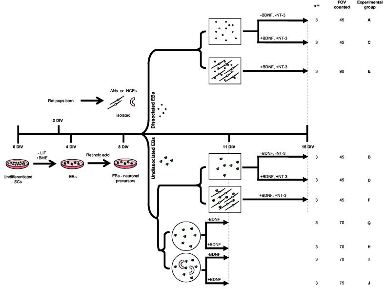
Figure 1.
Auditory co-culture models and control treatments
Stem cells (SCs) were co-cultured with rat post-natal day five auditory neurons or hair cell explants as described. Prior to co-culture, undifferentiated SCs were induced to form embryoid bodies via timed exposure to retinoic acid. Following eight days differentiation, SCs were placed into co-culture either dissociated or whole (illustrated schematically). The square wells represent treatments grown in auditory neuron media. Auditory neuron co-cultures and control treatments were maintained for 7 days in vitro. Circular wells represent hair cell explant co-cultures and relative controls, which were maintained for 3 days in vitro. Co-cultures and control treatments are distinguished pictorially. Following growth in vitro, all wells were fixed and processed for quantitative analysis. The number of times each experiment was repeated (n), the total fields of view (FOV) counted, and the experimental groups (A-J) are detailed on the far right had side of the diagram. DIV = days in vitro; SC = stem cell; LIF = leukemia inhibitory factor; BME = ß-mercaptoethanol; EBs = embryoid bodies; AN = auditory neuron; HCE = hair cell explant; BDNF = brain derived neurotrophic factor; NT3 = neurotrophin-3. Timeline is not to scale.

