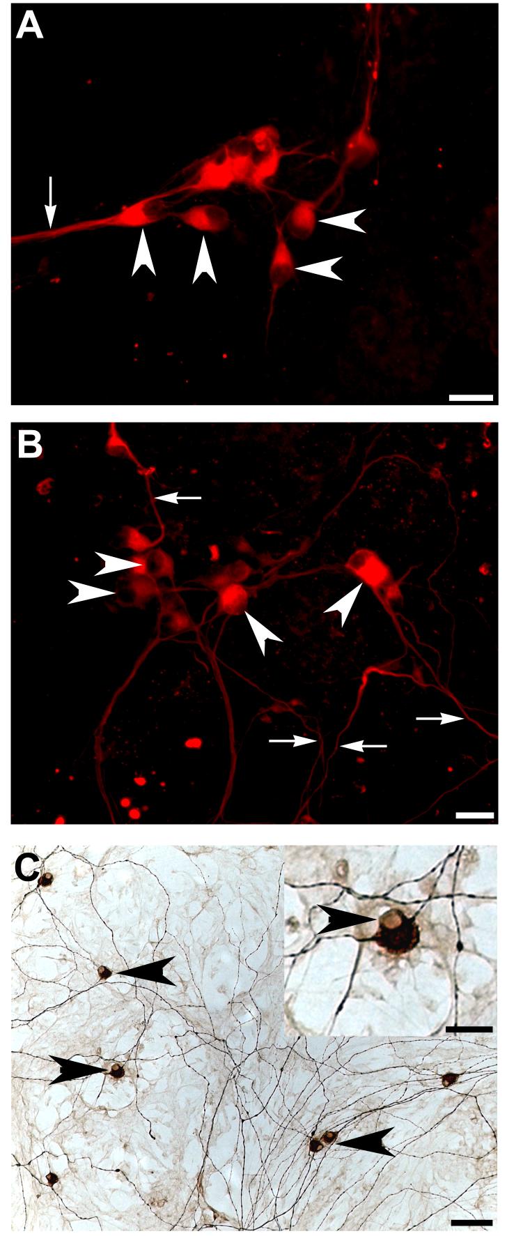
Figure 4.
Neurofilament positive bipolar cells were identified in hair cell explant co-cultures
Whole embryoid bodies were observed to differentiate into bipolar, neurofilament positive neuron-like cells when co-cultured with hair cell explants (arrowheads A,B). While some of these cells extended processes in the same direction (arrows A), others were observed growing in multiple directions (arrows B). Neurofilament positive cells displayed the characteristic bipolar morphology of early post-natal rat auditory neurons grown in vitro and immunolabelled with neurofilament protein (arrowheads, C). In particular, note the characteristic labelling around the nucleus (arrowhead, inset C). Scale bar = 20μm (A,B, inset C); 50 μm (C).

