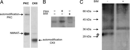
Fig. 1.
PKC-mediated phosphorylation of NMNAT-1. (A) Recombinant NMNAT-1 (500 ng) was incubated with PKC or casein kinase II (CKII) (10 units) and [γ-32P]ATP. Proteins were then separated by SDS/PAGE. The autoradiograph of the gel is shown. (B) NMNAT-1 (500 ng) was incubated with nuclear extracts (1 μg) in the presence of [γ-32P]ATP and phorbol myristate acetate (PMA) or BIM as indicated. Proteins were separated by SDS/PAGE, and gels were analyzed by autoradiography. (C) Human fibroblasts were incubated with [32P]orthophosphate for 8 h. Where indicated, BIM (1 μM) was added 1 h before cell lysis. After immunoprecipitation of endogenous NMNAT-1, proteins were separated by SDS/PAGE. Labeled proteins were visualized by autoradiography. The numbers on the left indicate the mobility of marker proteins. The asterisk shows the position of NMNAT-1.

