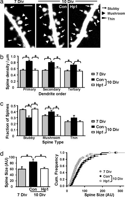
Fig. 3.
Knockdown of PSD-95 arrests spine morphological development. (a) Two-photon images of secondary apical dendrites at 7 Div, and at 10 Div after expressing control or effective shRNA for 3 days. (Scale bar: 2 μm.) (b) Hp1 prevented the increase in spine density (∗, P < 0.01). Primary dendrites: 0.55 ± 0.03 (7 Div), 0.84 ± 0.04, and 0.54 ± 0.05 spines per micrometer (Con and Hp1 at 10 Div). Secondary dendrites: 0.55 ± 0.02 (7 Div), 0.84 ± 0.04, and 0.62 ± 0.05 spines per micrometer (Con and Hp1 at 10 Div). Tertiary dendrites: 0.53 ± 0.02 (7 Div), 0.72 ± 0.05, and 0.52 ± 0.04 spines per micrometer (Con and Hp1 at 10 Div). No difference was observed between 7 Div and Hp1 at 10 Div (P > 0.13). (c) Hp1 prevented changes in spine type (∗, P < 0.05). Fraction of stubby spines: 0.43 ± 0.01 (7 Div), 0.30 ± 0.03, and 0.44 ± 0.02 (Con and Hp1 at 10 Div). Fraction of mushroom spines: 0.33 ± 0.02 (7 Div), 0.40 ± 0.02, and 0.32 ± 0.03 (Con and Hp1 at 10 Div). The fraction of thin spines did not change (P > 0.16). (d) Hp1 prevented spine size changes (∗, P < 0.04). Average spine sizes were 59.8 ± 7.9 (7 Div), 84.1 ± 9.7 (Con 10 Div), and 61.1 ± 4.7 (Hp1 10 Div; P = 0.44 vs. 7 Div) in arbitrary units (AU). (Right) Cumulative distributions of spine sizes. n = 6 cells per group.

